
[ad_1]
The cranial bone within the human physique performs crucial features, comparable to defending the mind and enabling the passage of the cranial nerves which might be important to physiological functioning. Important-sized cranial defects can disrupt each the bodily and psychological well-being of sufferers. Restoration of critical-sized cranial defects by cranioplasty is difficult for reconstructive surgeons, preferring to make use of autologous bone grafts. The acquisition of autologous bone requires extra surgical procedures concomitant with dangers comparable to free flap loss, an infection, deep venous thrombosis, and nerve damage. These limitations necessitate the event of options to autologous bone grafts for cranial defect restoration.
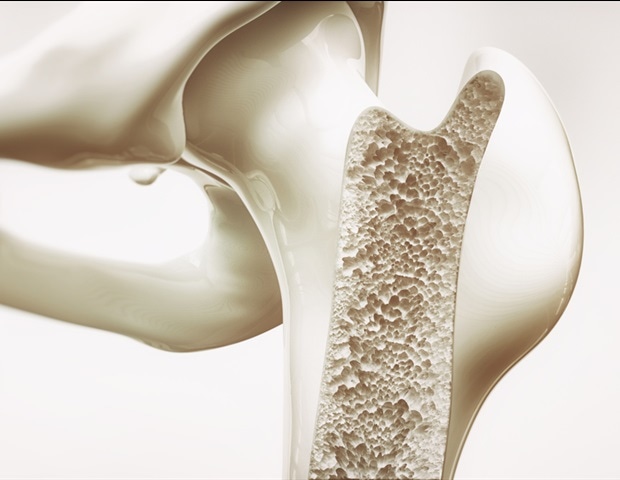
Biomaterials mimicking the composition and microstructure of pure bone are extensively acknowledged to be ultimate for bone defect regeneration The cranial bones are primarily composed of calcium phosphate, and they’re typical flat bones, that are typically skinny and broad with a flattened or curved floor. The flat bone has two outer compact tables made from cortical bone. The area between the 2 tables is known as the diploe, which consists of cancellous bone. Cortical bone has low porosity (5% to 10%) with interconnected tube-like pores, that are referred to as Haversian canals and Volkmann canals. The cancellous bone consists of irregular sponge-like trabeculae with a excessive particular floor space, and the imply floor curvature of the trabeculae floor is near zero. The architectural traits of cancellous bone are much like these of gyroid-type triply periodic minimal floor (TMPS) pore topology.
Impressed by the composition and structural options of cranial bones, scientists from South China College and Know-how developed two flat-bone-mimetic β-tricalcium phosphate bioceramic scaffolds (Gyr-Comp and Gyr-Tub) by high-precision vat photopolymerization-based 3D printing. Each scaffolds had two outer layers and an internal layer with gyroid pores mimicking the diploe construction. The outer layers of Gyr-Comp scaffolds simulated the low porosity of outer tables, whereas these of Gyr-Tub scaffolds mimicked the tubular pore construction within the tables of flat bones. The Gyr-Comp and Gyr-Tub scaffolds possessed increased compressive power and noticeably promoted in vitro cell proliferation, osteogenic differentiation and angiogenic actions in contrast with typical scaffolds with cross-hatch constructions. After implantation into rabbit cranial defects for 12 weeks, Gyr-Tub achieved the perfect repairing results by accelerating the technology of bone tissues and blood vessels. The Gyr-Tub scaffolds have excessive prospects for treating cranial bone defects in scientific purposes. This work offers a sophisticated technique to organize biomimetic biomaterials that match the structural and practical wants of efficacious bone regeneration.
Supply:
Journal reference:
Zhang, Y., He, F., Zhang, Q., Lu, H., Yan, S., & Shi, X. (2023). 3D-printed flat-bone-mimetic bioceramic scaffolds for cranial restoration. Analysis. doi.org/10.34133/analysis.0255.
[ad_2]